When Wilhelm Roentgen discovered X-rays in 1895, he opened the door to a new era of medicine. For the first time, doctors could see inside the human body without surgery. More than a century later, X-rays remain one of the most widely used imaging tools, and modern technology has made them safer, faster, and more precise than ever before.
At Iowa Radiology, we believe imaging is only the beginning. While X-rays are often the first step in diagnosis, their true value lies in the insights they provide–helping patients and providers make informed decisions about next steps in care.
A Brief History of X-rays
Roentgen’s discovery was revolutionary. Within months, X-rays were being used to detect bone fractures and locate bullets in injured soldiers. Marie Curie later advanced the field by developing mobile X-ray units for battlefield medicine during World War I. Over the decades, the technology continued to evolve, becoming faster, more accurate, and more accessible.
Today, X-rays are no longer limited to plain films of bones. They are integral to modern diagnostic imaging, used in procedures such as mammography, CT scans, and fluoroscopy. The technology that started more than 125 years ago still plays a central role in healthcare.
X-ray Safety
One of the most common questions patients ask is whether X-rays are safe. The answer is yes. While X-rays use ionizing radiation, the doses in modern exams are extremely low. For example:
- A chest X-ray exposes you to less radiation than the average person receives from natural background sources in 10 days.
- Mammograms use low-dose technology to minimize exposure while maximizing accuracy.
- CT scans use more radiation, but advances in technology have significantly reduced the dose required to produce clear images.
At Iowa Radiology, patient safety is always a priority. Our technologists and radiologists follow established guidelines to keep exposure “as low as reasonably achievable”, ensuring the benefits of imaging far outweigh any risks.
Should I Be Shielded During Imaging?
For many years, patients were given protective lead shields during X-rays. However, advances in technology and a better understanding of radiation have changed this practice. Shields can sometimes block important anatomy or even increase exposure by interfering with the imaging equipment’s automatic settings. Today, professional organizations recommend against routine shielding for most exams.
At Iowa Radiology, we follow the latest evidence-based practices, prioritizing safety while making sure images are as accurate as possible.
The Role of X-rays in Cancer Screening and Early Detection
X-rays aren’t just for broken bones. They remain central to cancer detection:
- Mammography uses X-ray technology to detect breast cancer at its earliest stages. With 3D mammography, detection is more accurate than ever.
- Low-dose CT (LDCT) for lung cancer screening relies on X-ray technology to capture highly detailed images of the lungs with minimal exposure.
- Virtual colonoscopy uses CT scans (powered by X-rays) to screen for colorectal cancer without sedation.
Each of these applications highlights how X-rays continue to save lives by finding cancer before symptoms appear.
What Can a Chest X-ray Tell Me?
Chest X-rays are one of the most common imaging tests, often ordered as part of routine care or when symptoms such as cough, shortness of breath, or chest pain arise. A chest X-ray can show:
- The size and shape of the heart.
- Evidence of pneumonia, bronchitis, or other lung infections.
- Chronic lung conditions like emphysema or COPD.
- Abnormal growths or fluid around the lungs.
While a chest X-ray may not provide all the answers, it often serves as an important starting point that guides further testing.
Mammograms and Radiation
Some women worry about radiation exposure from mammograms. It’s important to know that modern mammography uses very low doses of radiation. At Iowa Radiology, we use 3D mammography, which not only improves cancer detection, especially in women with dense breasts, but also minimizes unnecessary callbacks and repeat imaging.
The small amount of radiation used in a mammogram is considered safe, and the benefit of detecting breast cancer early far outweighs the risk.
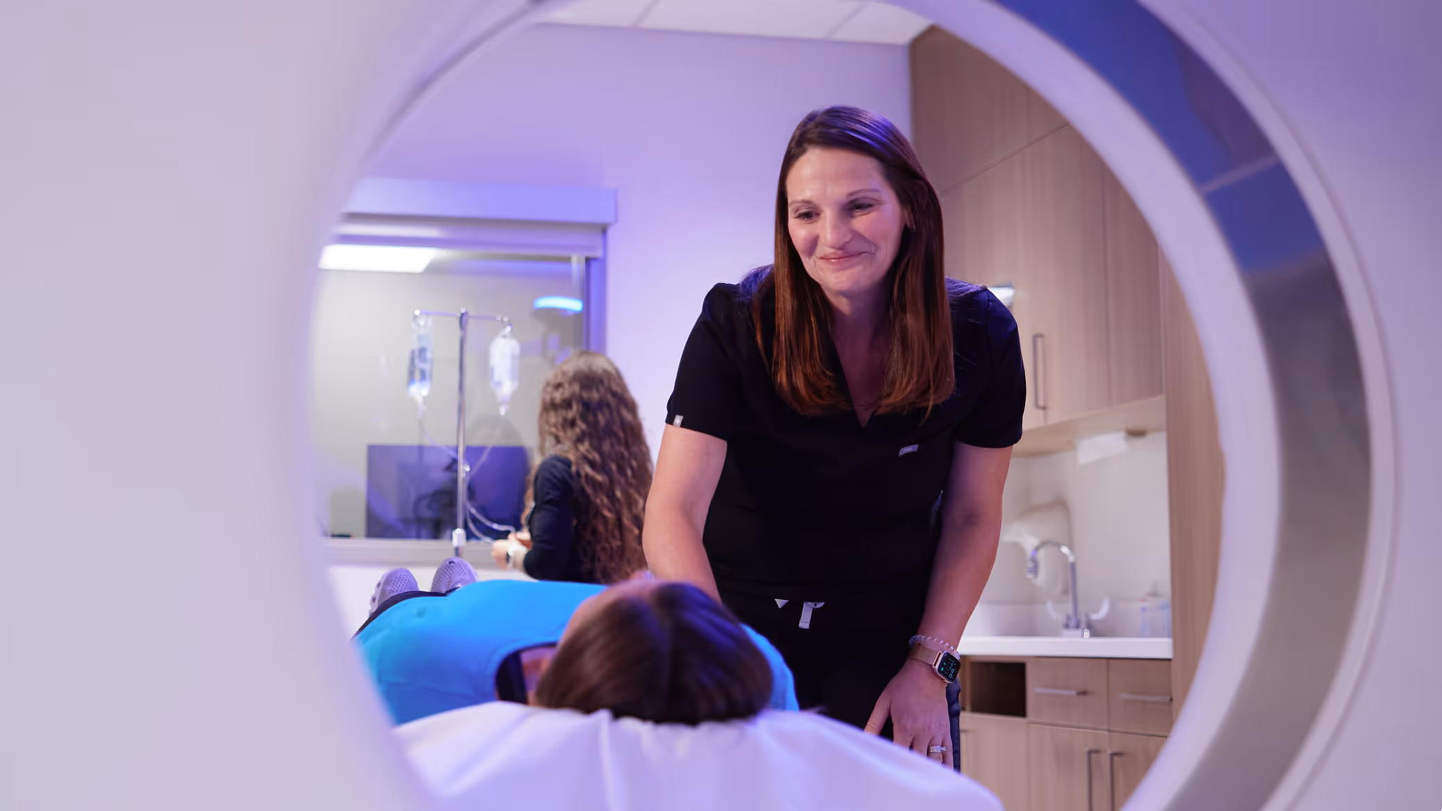
The Iowa Radiology Difference in X-ray Imaging
While X-rays are often thought of as “routine,” their interpretation requires expertise. At Iowa Radiology, patients benefit from:
- Advanced Technology. We use digital X-ray systems that provide faster results with lower exposure.
- Experienced Radiologists. Our team has expertise across multiple areas, ensuring accurate readings that guide care.
- Clear Communication. Results are shared promptly with your provider so next steps can be taken without delay.
- Patient-Centered Care. We explain the process, answer questions, and ensure every patient feels informed and comfortable.
Old Technology, New Impact
X-rays may be the oldest imaging technology, but they continue to make a modern impact. From routine chest X-rays to advanced cancer screenings, X-ray technology provides clarity that shapes patient care every day.
At Iowa Radiology, we’re proud to carry forward this legacy with the latest technology and a commitment to delivering insights that guide care. If your provider recommends an X-ray or another imaging test, you can trust that our team will deliver results that do more than capture an image; they’ll help chart the next step in your health journey.